Cathodoluminescence Imaging of Organic Fluorophores and Fluorescently Labeled Cells
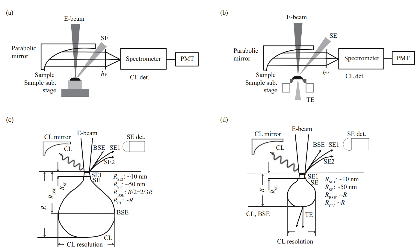
The correlative cathodoluminescence (CL) imaging is a high-resolution fluorescence imaging technique using electron beam as excitation source. However, the sensitivity of biomaterials to electron irradiation limits the wide application of CL technique in life science. In order to study and develop the application of CL technology in biological samples, this paper aims to deeply understand the excitation characteristics of organic fluorophore by electron source by exploring the structural damage of carbon substrate, degradation of organic groups and fluorescence quenching caused by electron irradiation.Methods We used a CL and electron microscopy (CCLEM) technique in a field emission scanning electron microscope (SEM) to observe organic fluorophores and fluorescently labeled cells.Results We studied excitation and emitting characteristics of organic fluorophores by electron beam. Under the irradiation of low energy (2.5-5 keV) and low current (~10 pA), the fluorescent microbeads had the stronger and stable CL emission, the resolution of CL image reached ~30 nm. After 12 min irradiation, fluorescence intensity of microbeads decreased by 25%, and CL image still maintained acceptable fluorescence intensity and adequate signal to noise ratio. Further, fluorescently labeled subcellular structures were identified from cell surface to a certain depth inside the cell. The nuclei and organelles were well observed with a bulk-sample imaging setup, while membrane proteins, that are widely localized on the cell surface, were clearly identified with a home-made imaging setup of thin-sample.Conclusion Results provide data and technical support for the application of CCLEM in the study of biological structures. CCLEM bioimaging can also be used as an important supplement to correlative light and electron microscopy (CLEM) technique.
Article Link: 有机荧光团及荧光标记细胞的阴极荧光成像研究 (pibb.ac.cn)
附件下载: